咨询热线: 400-670-9909
联系我们
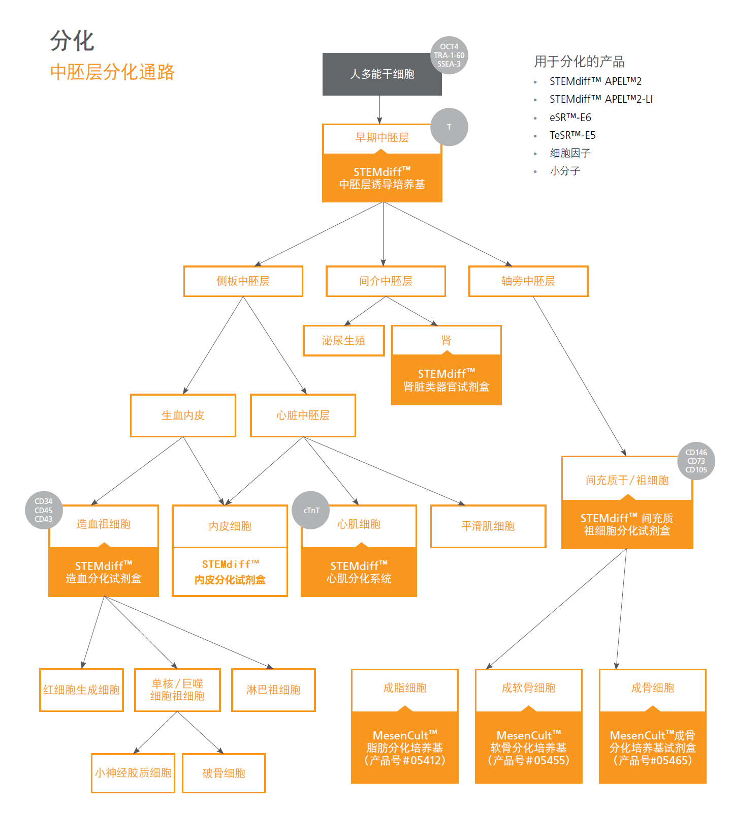
一、hPSC中胚层分化通路及产品一览表
二、新品推荐
STEMCELL 内皮细胞分化培养基:STEMdiff Endothelial Differentiation Kit(#08005),该培养基是市面上首套优化用于hPSC分化为内皮细胞并进行扩增的培养基。对于想进一步扩增hPSC来源的内皮细胞的客户,可以单独购买STEMdiff内皮细胞扩增培养基试剂盒:STEMdiff Endothelial Expansion Medium Kit (#08007)。

三、产品规格
| STEMdiff Endothelial Differentiation Kit | Cat #08005 | 规格 |
| STEMdiff Endothelial Induction Medium | #08006 | 100 mL |
| STEMdiff Endothelial Expansion Basal Medium | #08008 | 120 mL |
| STEMdiff Endothelial Expansion (5X) Supplement | #08009 | 30 mL |
| Animal Component-Free Cell Attachment Substrate | #07130 | 1 mL |
| STEMdiff Endothelial Expansion Medium Kit | Cat #08007 | 规格 |
| STEMdiff Endothelial Expansion Basal Medium | #08008 | 120 mL |
| STEMdiff Endothelial Expansion (5X) Supplement | #08009 | 30 mL |
四、产品优势
STEMdiff Endothelial Differentiation Kit 经过优化,可有效地将hPSC分化为内皮细胞,并在多个hPSC细胞系中均具有很好的稳定性。
内皮细胞诱导后,在STEMdiff Endothelial Expansion Medium中培养hPSC衍生的内皮细胞,通过P1-P2代可以产生高纯度的内皮细胞,并且不需要DIY方案中所需的细胞富集步骤。
STEMdiff Endothelial Expansion Medium 支持高效的hPSC衍生内皮细胞扩增,优于使用含FBS的培养基配方获得的内皮细胞扩增效率。
使用STEMdiff Endothelial Expansion Medium扩增的内皮细胞保留了内皮细胞的表型,并在传代后也保持内皮细胞的表型和功能。
五、部分数据
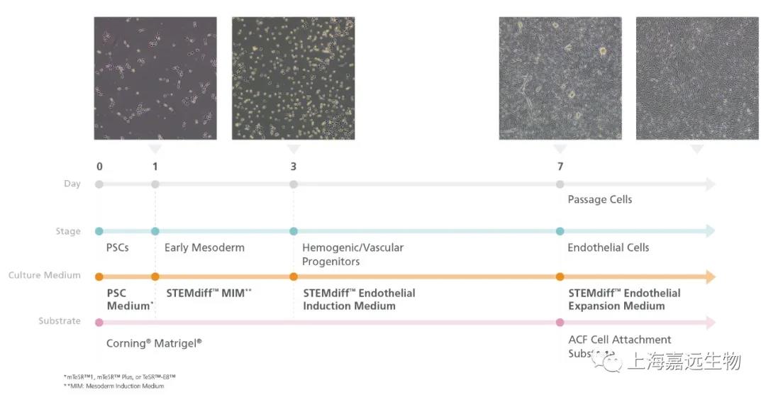
Figure 1. Schematic Workflow of Endothelial Induction Using the STEMdiff™ Endothelial Kit
In Phase 1, human embryonic stem (ES) or induced pluripotent stem (iPS) cells are cultured in a TeSR™ maintenance medium (mTeSR™ Plus, mTeSR™1, or TeSR™-E8™). On Day 1 (Phase 2) of the protocol, cells are ready for induction into early mesoderm progenitor cells by replacing TeSR™ medium with STEMdiff™ Mesodermal Induction Medium (MIM). By Day 3 (Phase 3), STEMdiff™ Mesoderm Induction Medium is replaced with STEMdiff™ Endothelial Induction Medium to derive endothelial cells. On Day 7, cells are passaged 5 - 6 times onto cultureware pre-coated with Animal Component-Free Cell Attachment Substrate in STEMdiff™ Endothelial Expansion Medium (Phase 4).

Figure 2. A Representative Flow Cytometric Analysis of Endothelial Marker Expression in hPSC-Derived Endothelial Cells
Human pluripotent stem cell (hPSC; H9 cell line)-derived endothelial cells were obtained at Day 7 using STEMdiff™ Endothelial Induction Medium. Greater than 85% of the cells were CD34+ and had high levels of CD31 and CD144 expression. With subsequent passages, the proportion of cells expressing endothelial markers (CD34+, CD31, and CD144) increased up to passage 5.
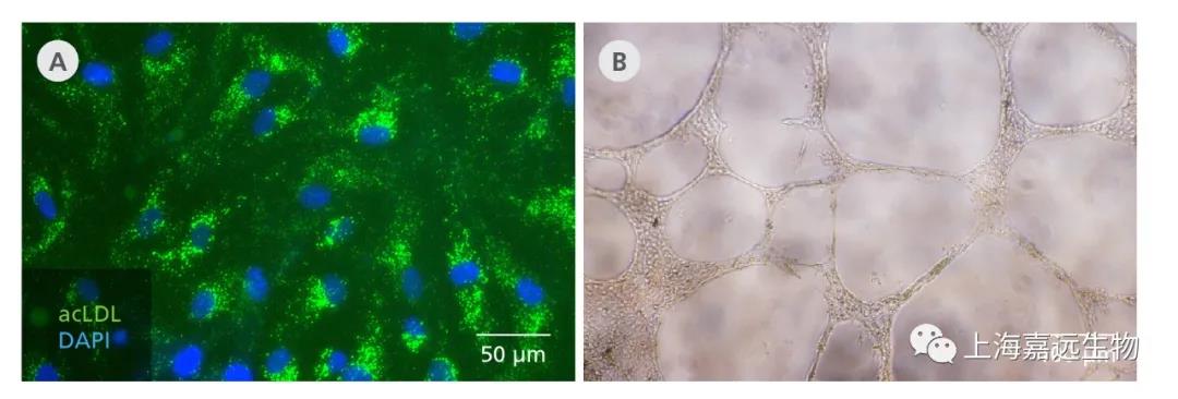
Figure 3. STEMdiff™ Endothelial Differentiation Kit Generates Functional hPSC-Derived Endothelial Cells
Endothelial cells generated from hPSCs (F016 cell line) using the STEMdiff™ Endothelial Differentiation Kit take up acetylated LDL when plated at 10,000 cells/cm2. Cells are able to form tubular networks in vitro in a tube formation assay when plated at 20,000 cells/well in a 96 well-plate for 24 hrs.
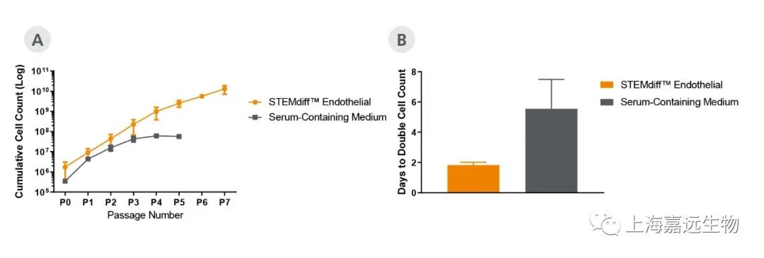
Figure 4. Endothelial Cells Expand Faster in STEMdiff™ Endothelial Expansion Medium Compared to Serum-Containing Medium
STEMdiff™ Endothelial Expansion Medium (A) sustains expansion rate in later passages and leads to (B) superior expansion of hPSC (C1 cell line)-derived endothelial cells when compared to serum-containing medium.
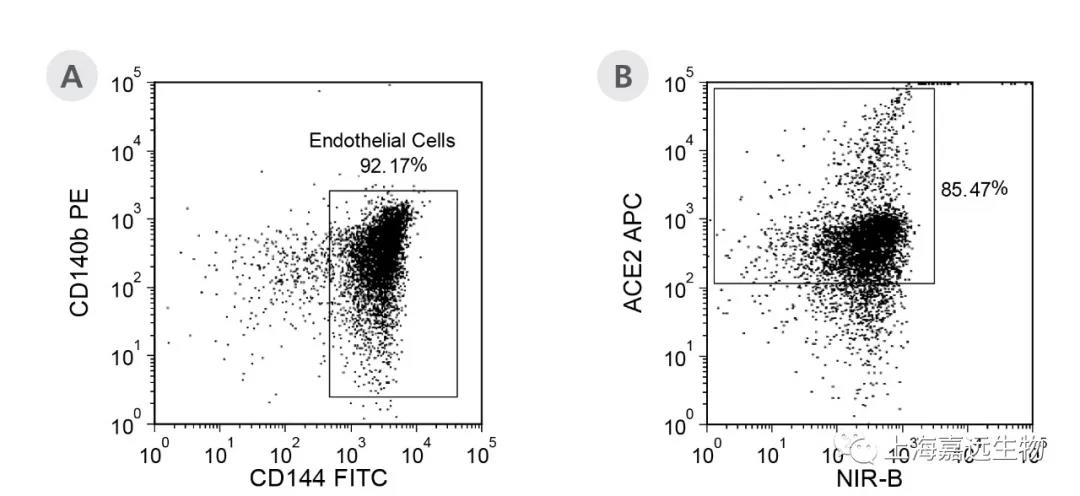
Figure 5. hPSC-Derived Endothelial Cells Generated Using the STEMdiff™ Endothelial Differentiation Kit Express High Levels of ACE2
(A) hPSC (C1 cell line)-derived endothelial cells were generated using the STEMdiff™ Endothelial Differentiation Kit and expanded in STEMdiff™ Endothelial Expansion Medium for 6 passages at 10,000 cells/cm2. (B) The cells were then analyzed for expression of angiotensin-converting enzyme 2 (ACE2). 85% of cells expressed high levels of ACE2.


 沪公网安备 31011202004777号 技术支持:赤云网络